Researchers at the Korea Advanced Institute of Science and Technology have identified a previously unrecognised mechanism by which a checkpoint inhibitor drug prompts a B cell and antibody response in lymph nodes deep within the neck, a discovery that may open a novel avenue to treat glioblastoma and other difficult to treat brain tumours
Researchers have uncovered a clue as to why immune checkpoint inhibitors – cancer therapies that release the immune ‘brakes’ exploited by tumours to evade attack – have shown limited efficacy against some brain tumours. A team at the Korea Advanced Institute of Science and Technology (KAIST) have found that B‑cell and antibody responses initiated in tumour‑draining lymph nodes – rather than T cells alone – are critical to the antitumour effects of anti‑cytotoxic T‑lymphocyte‑associated protein 4 (anti‑CTLA‑4) therapy, a finding that opens up a novel pathway to treat intractable brain tumours.
KAIST have said that a research team led by Professor Heung Kyu Lee of the department of biological sciences has identified a previously unrecognised immune mechanism through which anti‑CTLA‑4 – a type of immune checkpoint inhibitor – promotes B‑cell responses in tumour‑draining lymph nodes thereby helping the immune system to attack brain tumours.
Glioblastoma is among the most aggressive malignant brain tumours, with frequent recurrence and a poor prognosis even after surgery and radiotherapy. Immune checkpoint inhibitors, which restore the ability of immune cells to attack cancer cells, have produced substantial therapeutic benefits in various cancers. However their effectiveness against glioblastoma has remained limited because of the highly immunosuppressive environment that surrounds the tumour.
Researchers have traditionally regarded T cells as the primary target of checkpoint inhibitors. Meanwhile B cells are well known for their capacity to produce antibodies following infection or vaccination but their role in brain tumour immunotherapy had remained largely unexplored. The KAIST team therefore set out to establish whether anti‑CTLA‑4 could influence B‑cell responses as well as T‑cell responses.
The findings challenged the prevailing T‑cell‑centred view of how these drugs work. In mouse models of glioma, anti‑CTLA‑4 treatment reduced tumour burden and significantly prolonged survival. These therapeutic effects were, however, largely lost in mice that lacked B cells, a result that indicated B cells were required for anti‑CTLA‑4 treatment to be effective in these models.
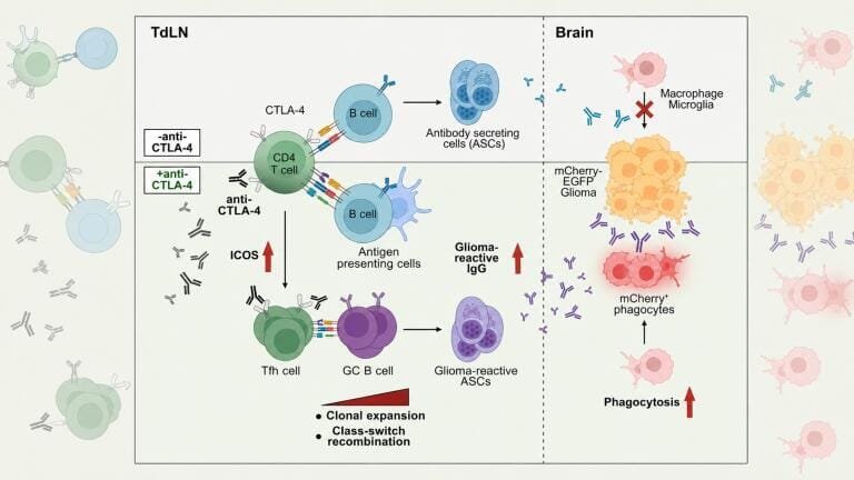
The team also established where B cells play their key role. Rather than being prominent in the brain itself, where the tumour cells were located, the response increased markedly in the deep cervical lymph nodes sitting deep within the neck and receiving lymphatic drainage from the brain. Germinal centre B cells and T follicular helper cells, both important to antibody formation, increased together within these lymph nodes, a change accompanied by a rise in immunoglobulin G (IgG) responses. IgG is a major class of antibody able to recognise cancer cells as targets and to help immune cells eliminate them.
The resulting IgG antibodies bound to the surface of glioma cells, helping macrophages – immune cells that engulf foreign substances and cancer cells – to remove the tumour cells more effectively.
The research team went on to examine this process directly in living tissue. Using a specialised dual‑reporter glioma model that expressed the red fluorescent protein mCherry and the green fluorescent protein, the researchers were able to visualise tumour‑infiltrating phagocytes actively engulfing glioma cells following anti‑CTLA‑4 treatment.
The study provides the first functional evidence that B‑cell immune responses can be a key factor in determining the effectiveness of immunotherapy for hard‑to‑treat brain tumours. It also expands the conventional T‑cell‑centred framework of cancer immunotherapy by showing that treatment efficacy can be shaped strongly by immune responses that originate not only within the tumour but also in tumour‑draining lymph nodes some distance from it.
As the results derive from mouse models rather than human trials, any translation into a treatment for people with glioblastoma or other brain tumours would still require substantial further research, including safety and efficacy testing in humans.
Dr. Yumin Kim, a postdoctoral researcher in the KAIST department of biological sciences, served as first author of the study, with Professor Heung Kyu Lee as corresponding author. Professor Ji Eun Oh of the KAIST graduate school of medical science and engineering also contributed to the research.
For further reading please visit: 10.1126/sciimmunol.adz2494
Picture caption:
Figure 1. Proposed model. In glioma-bearing mice, anti-CTLA-4 treatment increases inducible T cell costimulator (ICOS) expression on CD4 T cells and expands T follicular helper (TFH) cells in tumour-draining dcLNs. Enhanced TFH responses promote the expansion of germinal centre (GC) B cells, leading to clonal expansion and class switching. This induces glioma-reactive IgG production, which binds glioma cells and enhances phagocytosis by tumour-infiltrating myeloid cells, contributing to glioma control. Graphics created with BioRender. Credit: KAIST