Probing flash-frozen biological samples with an electron beam has been a game changer for researchers examining the details of biological structures. The technique—cryogenic electron microscopy, known as cryo-EM—has exploded in popularity in the past 15 years. It earned some of its inventors the 2017 Nobel Prize in Chemistry and was used in 2020 to analyze the structure of the COVID-19 virus’s spike protein (see the 2020 PT story “World’s physics instruments turn their focus to COVID-19 ”), an achievement that enabled development of targeted vaccines.
Still, most proteins in the human body are too small to be effectively imaged with cryo-EM. The primary challenge is that biological materials refract electrons only weakly, so the images are inherently low contrast. And because electron beams damage biological samples, lengthening exposure times isn’t a viable solution.
A new advance by a team led by physicist Holger Müller at the University of California, Berkeley, improves the resolution of cryo-EM and offers promise for clearer images of the cellular world. The researchers use the highest-intensity continuous-wave laser ever demonstrated—about 400 GW/cm2, 100 million times as bright as the Sun’s surface—to phase shift the electron beam.
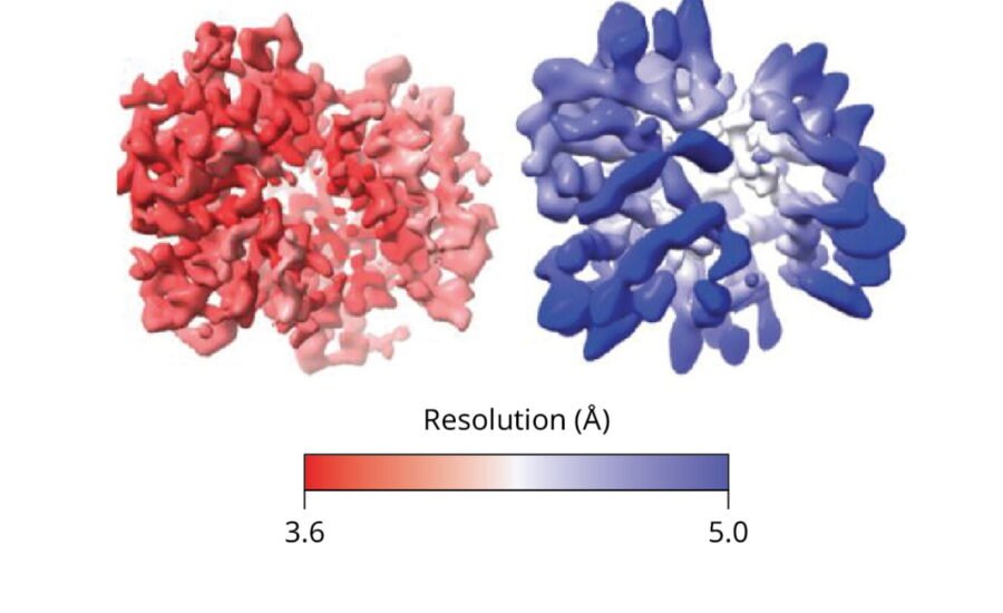
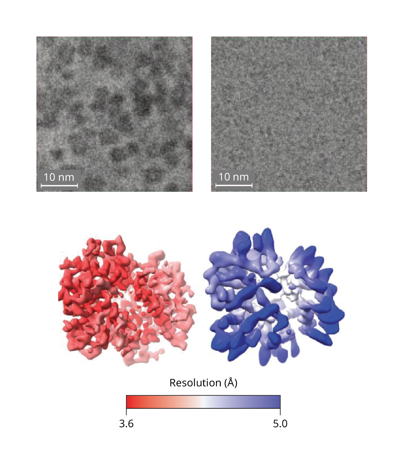
That phase shift produces cryo-EM images with heightened contrast and produces higher-resolution models of proteins, such as the hemoglobin molecule shown in figure
1
.
Figure 1.

A micrograph of hemoglobins collected with a laser phase plate–fitted electron microscope (top left) has better contrast than one collected without the plate (top right). Thousands of protein images are analyzed to produce a structural model of hemoglobin. The higher-contrast images yield a structural model (bottom left) that captures more details of hemoglobin’s shape than does a model produced from lower-contrast images (bottom right).
(Figure adapted from ref.
.)
Fuzzy details
In typical cryo-EM, a combination of strategies is used to extract useful information from low-contrast images. One counterintuitive tactic is to collect intentionally defocused images to create contrast. Consider a blurry photocopy of a book page on which each line of text appears as a fuzzy gray line. Details of individual words and letters are sacrificed, but the lines stand out more. Without defocusing, cryo-EM images have essentially no contrast. (For more on the methodology of cryo-EM, see Bob Glaeser’s 2008 PT article, “Cryo-electron microscopy of biological nanostructures .”) Researchers using cryo-EM collect up to hundreds of thousands of defocused images of molecules and then process them with algorithms that analyze proteins’ shapes and orientations to produce a single structural model.
Though biological samples also produce weak scattering and low contrast in light-based microscopy, an effective technique to deal with that problem was deduced in the 1930s: Phase shift the direct light—the portion that has not been diffracted while passing through the sample—by 90°. The phase-shifted light waves produce constructive and destructive interference with the scattered waves, which enhances the contrast of the subsequent image without the need for defocusing. (Frits Zernike was awarded the 1953 Nobel Prize in Physics for the phase-contrast method.) Though it was quickly recognized that the same approach could benefit electron microscopy, phase shifting electrons has not been as straightforward as phase shifting light.
Before turning to lasers, researchers had explored other methods to phase shift electron beams and improve cryo-EM contrast. The most successful phase-shifting approach has been the Volta phase plate, a heated thin film of amorphous carbon. Interaction between the plate and the unscattered electron beam produces a voltage potential that phase shifts the beam. Though the Volta phase plate has succeeded at producing some of the best cryo-EM images to date, it also causes signal loss and has thus not yielded consistent improvements in resolution.
Laser focus
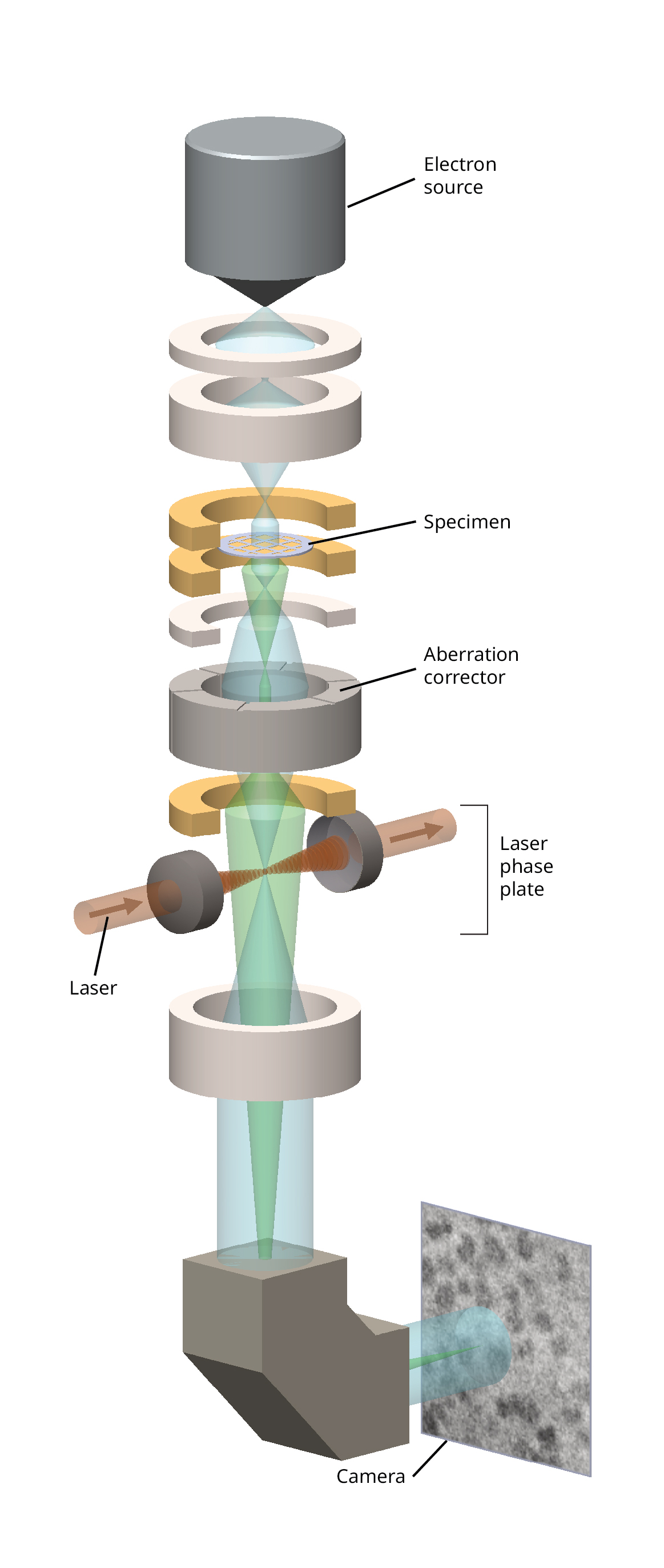
Müller and colleagues proposed in 2010 that passing the electron beam through a powerful laser, called the laser phase plate and illustrated in figure
2
, might do the trick.
Despite its name, there is no physical plate involved in the laser phase plate. Electrons entering the electric field of the laser experience a repulsive potential that shifts the phase of the electron wave function by an amount that is predictably related to the laser’s power and wavelength. To create a continuous-wave laser with the necessary 80 kW power, the researchers would need to bounce it back and forth between two curved mirrors about 10 000 times. The mirrors form a resonant cavity that amplifies the laser power with each traverse. The cavity also focuses the beam down to a diameter of about 7 µm, the width of a red blood cell, to yield the intensity of 400 GW/cm2.
Figure 2.

By adding an ultrabright laser to an electron microscope, researchers improved the contrast of biological images collected with the microscope and were able to see more details in the structure of small proteins.
(Figure adapted from ref.
.)
In early attempts, the mirrors got too hot and started to change shape when the laser power reached about 10 kW. The researchers changed the mirror material to a titania silicate glass with a low thermal expansion coefficient and achieved about 30 kW. To further improve the laser power, the researchers had to secure the mirrors so that the distance between them would remain at an exact multiple of half the laser wavelength.
Finally, around 2020, the laser was working as needed, and the team began collecting cryo-EM images. Though the contrast looked much better, the postprocessing resolution didn’t seem to improve. “We had a microscope that produced gorgeous-looking images. We were so happy,” says Müller. “But then you feed the images into the computer and say, ‘Tell me what’s the resolution from these images,’ and the computer says that it’s about the same as without the laser.”
To add the laser to the microscope, the researchers also had to add two more electron lenses, but those were introducing aberrations that negated the improvements from the laser. By upgrading to a state-of-the-art electron microscope that has an electron beam with a narrower energy spread and an aberration corrector, the benefits of the laser phase plate finally came into view.
The next phase
For their first analysis of images from the laser phase plate–fitted microscope, which is shown in figure
3
, Müller and colleagues used otherwise standard procedures for collecting cryo-EM images, including defocusing, so they could directly compare images collected with and without the plate. With a diameter of about 55 Å, hemoglobin molecules are on the smaller end of molecules that can be imaged with cryo-EM. By using the laser phase plate, the researchers enhanced the resolution of the molecule’s structural model—the largest improvement was from 4.46 Å down to 3.09 Å. The heightened contrast from the laser phase plate also means that defocusing should no longer be necessary for future data collection. The team plans to collect in-focus images that should reveal even finer details of protein structures.
The laser phase plate “makes a much larger pool of proteins available to study by cryo-EM, which is important because the majority of proteins in human cells are very small,” says Mohammed Kaplan, a microbiologist at the University of Chicago. He says the method should be especially useful for cryoelectron tomography, in which intact cells are imaged at multiple angles to produce 3D volumes that show proteins in their cellular environment.
Figure 3.

Holger Müller in front of the laser phase plate–fitted electron microscope.
(Photo courtesy of Robert Sanders/UC Berkeley.)
Another benefit of the laser phase plate is that frames collected at the beginning of a sample analysis should become more useful. When cryo-EM samples are prepared, they are plunged into liquid ethane to shock-freeze them. That creates a vitreous phase of ice that locks tension into the sample. Energy deposited by the electron beam releases the tension, and samples start to move erratically. As a result, the amount of noise in the earliest frames is high, and the images are discarded. The improved signal-to-noise ratio of the new technique, however, should make the early frames usable.
Müller got the funding to upgrade his lab’s electron microscope from the nonprofit research organization Biohub and has worked with researchers there to develop another version of the laser phase plate. That one, built at Biohub’s imaging lab, uses two crossing lasers at half power each and also significantly improves resolution, the researchers report in a bioRxiv preprint.
Both research teams are working with Thermo Fisher Scientific to commercialize the technology. Any ultimate product would probably require that researchers have a state-of-the-art electron microscope similar to the ones in Müller’s lab and at Biohub.
Beyond its value for cryo-EM, the ultrabright laser could have other uses. Müller’s lab is looking into using it as an optical trap that is compatible with a wider range of molecules than are current trapping methods.